A Diagnostic Challenge
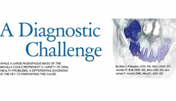
This case report is of a 15-year-old boy who presented for orthodontic treatment at a military base dental clinic. Upon radiographic examination, a large radiopaque mass was noted in the patient’s right maxilla (Figure 1).Computed tomography scans revealed that the mass was encroaching upon the posterior wall of the maxillary sinus (Figure 2 to Figure 4). The maxillary right second and third molars were impacted and associated with the lesion (Figure 5). The patient was asymptomatic. He had no sensation of fullness, history of swelling, or visual disturbance. Previous radiographs were unavailable for review. Several diagnoses are possible for a large radiopaque mass of the maxilla, including ameloblastic fibro-odontoma (AFO), compound odontoma, complex odontoma, ossifying fibroma, and cementoblastoma. A differential diagnosis is necessary to determine the correct cause of this lesion.
* References can be found in the original article via the link below.
Read Article


Responses