A Closer Look at Gingival Recession

By Keerthana Satheesh, DDS, MS
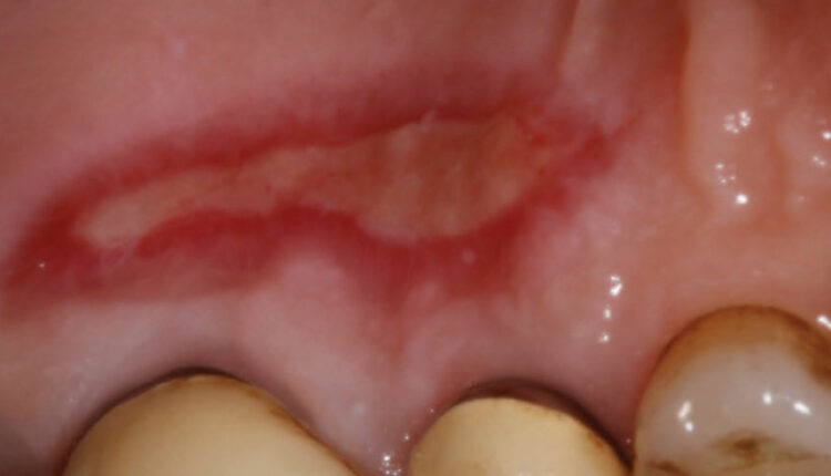
Gingival recession is associated with oral exposure of the root surface due to a displacement of the gingival margin apical to the cementoenamel junction.1 This is a fairly common clinical condition, and research indicates it presents in at least one or more tooth surfaces in 23% of United States adults between the ages of 30 and 90.2 According to the classification system developed by the American Academy of Periodontology (AAP), mucogingival deformities and conditions around teeth and edentulous ridges include gingival/mucosal tissue recession, lack of keratinized gingiva, decreased vestibular depth, aberrant frenal pull/muscle position, gingival excess, and abnormal color.3 While this article will focus on key aspects of gingival recession, it will not discuss decreased vestibular depth, gingival excess, or abnormal color.
* References can be found in the original article via the link below.
Read Article


Responses