The Safe and Effective Use of Cone-Beam Computed Tomography
By Brigette R. Cooper, RDH, MS, and Trisha Krenik-Matejcek, RDH, MS
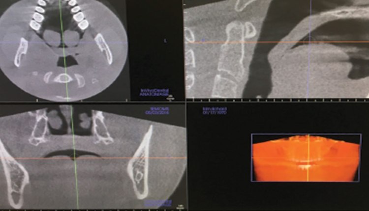
Acquiring high-quality radiographic images is essential to rendering accurate oral diagnosis and treatment. An important advancement in digital radiography is cone-beam computed tomography (CBCT). A radiographic imaging method, CBCT provides accurate, three-dimensional (3-D) imaging of hard tissue structures. CBCT is important to incorporate into specialized dental settings, as it provides a higher standard of care for patients.1 Although two-dimensional (2-D) images have provided diagnostic information in dentistry for many years, they have limitations—such as magnification, distortion, and superimposition of anatomical details—that can lead to misrepresentation of structures.1 As CBCT becomes more commonly used in dentistry, oral health professionals should become familiar with the diagnostic capabilities of this 3-D technology.
* References can be found in the original article via the link below.
Read Article


Responses